Blood Under a Microscope
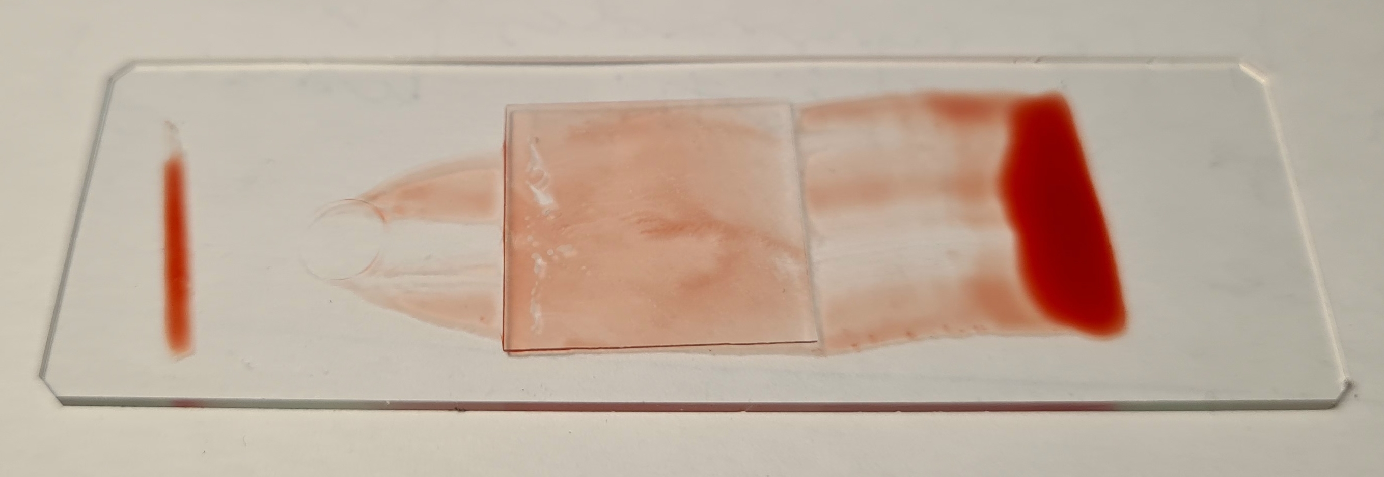
Getting a cut from a freshly sharpened knife was a good occasion to inspect blood under a microscope (one more time).

Here's a special calibration slide that can be used to measure objects visible under the microscope:
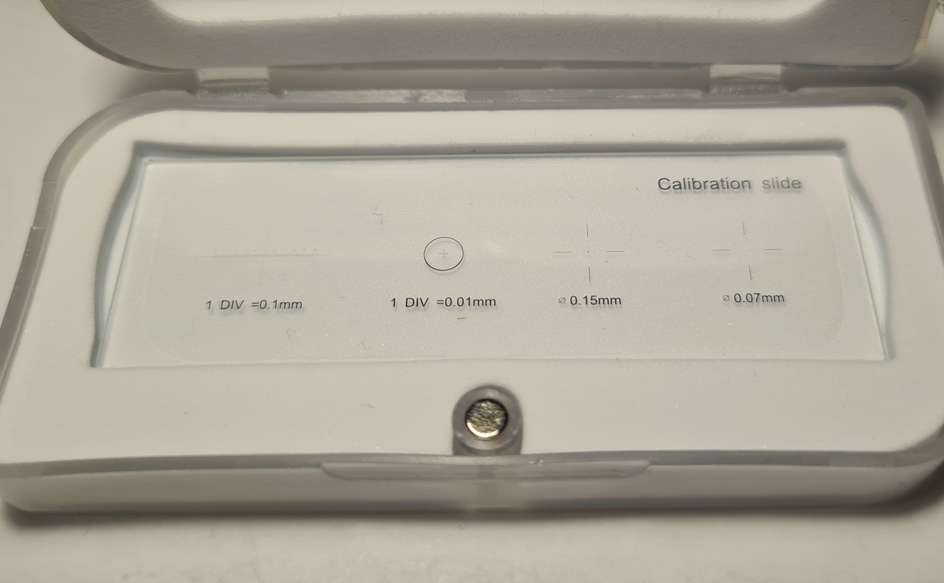
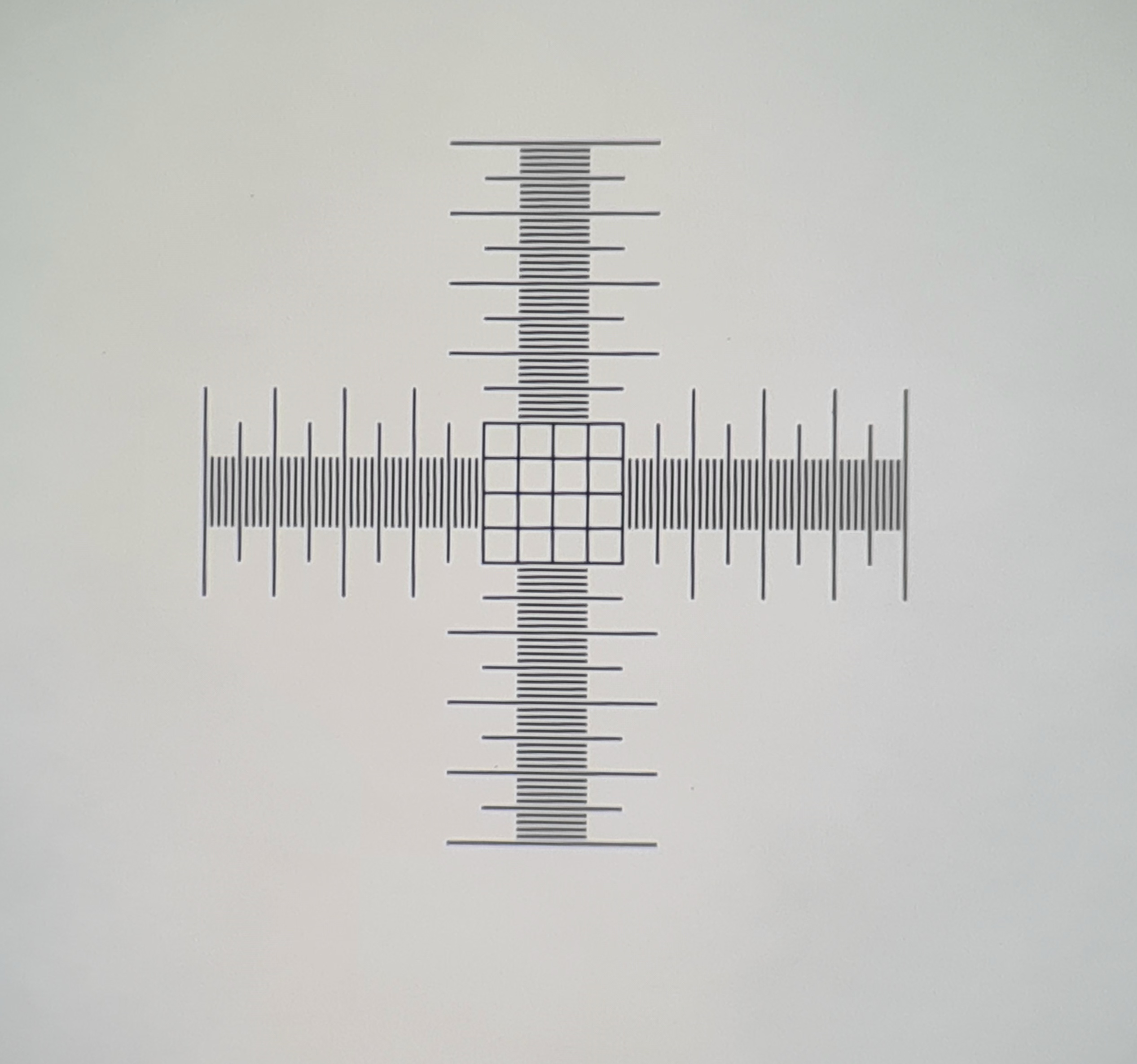
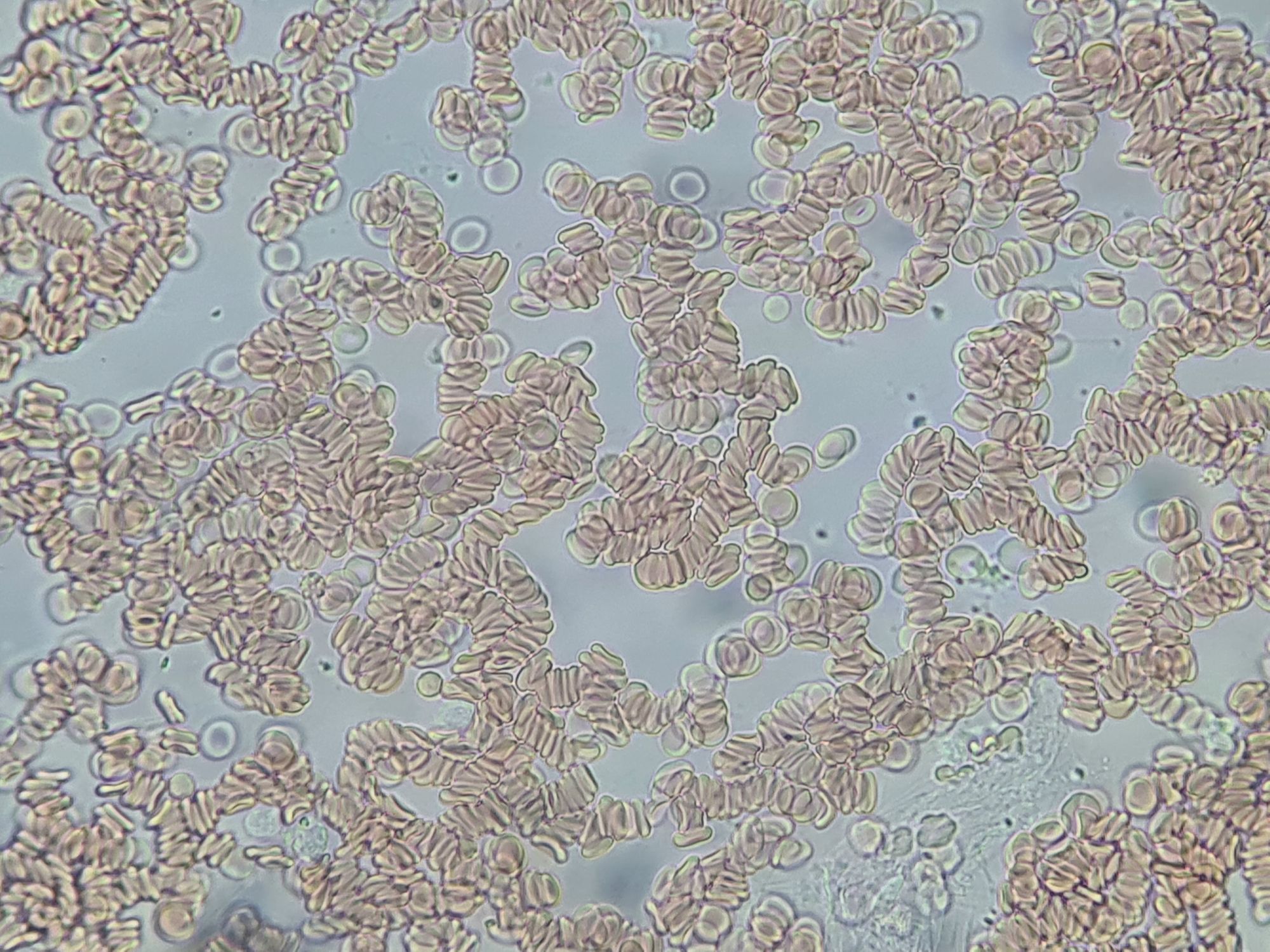
We can use this to measure size of the red blood cell by superimposing calibration image and red blood cells. Assuming centres as divisions we can roughly tell that a red blood cell is around 0,7 × 0,01 mm = 7 μm. Properly measured size is between 7,5 - 8,7 μm.
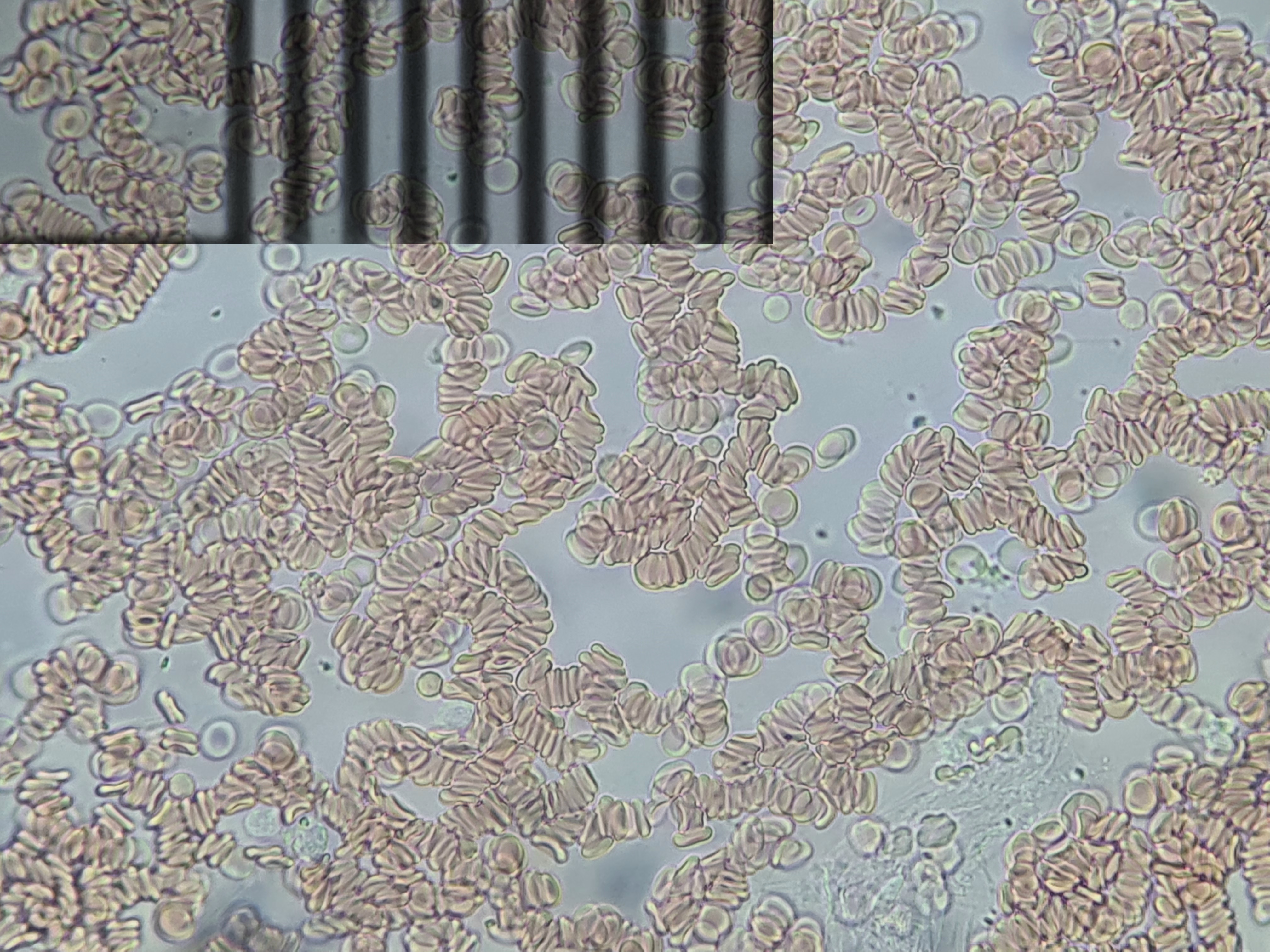
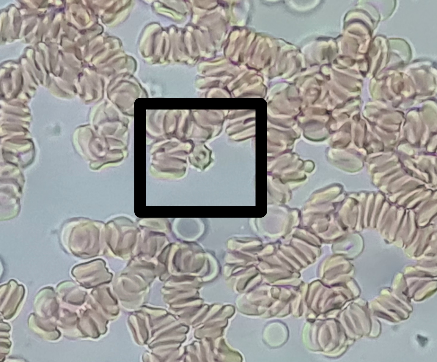
Without staining other cells can be seen, in black box is an activated platelet (caused by the cut).